Fetal Echocardiograms
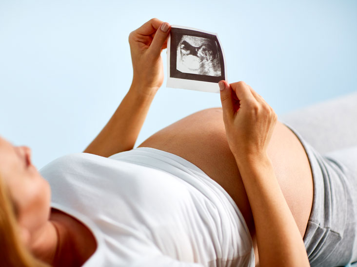
Fetal echocardiograms are types of ultrasound tests that reveal the physical structure and functioning of the unborn child’s heart. A pregnancy test of this importance is conducted between weeks 18 and 24 of the second trimester. The fetal echocardiogram has given many good results during pregnancy over the years. It is perfectly understandable that patients might get worried about their unborn child and want to know its health condition, so this test was created just for that purpose. By reflecting sound waves off the fetus’ heart, one can determine the structure of the child. Let’s dive into the article shared by Dr. Mamta Phogat – the best Fetal Medicine Specialist in Faridabad to know in detail.
Fetal echocardiograms are needed when?
A fetal medicine specialist in Faridabad will examine the patient and recommend the fetal echocardiogram test if any of the high-risk pregnancy symptoms have been observed. For most pregnant patients, a basic ultrasound will show the development of all four chambers of the unborn baby’s heart. A fetal echocardiogram is needed in the following circumstances:
- If the unborn child is at risk of abnormal heart and brain development.
- If there is a family history of heart disease.
- In the past, the patient has delivered a baby with a heart condition.
- During pregnancy, the patient has used drugs or alcohol.
- If the women are exposed to medication that causes heart defects.
Fetal echocardiograms are performed in what manner?
fetal echocardiography is a test performed by a well-trained special ultrasound sonographer. Patients who are found at fault for any of the above risks should be cautious and immediately consider fetal echocardiography. Fetal echocardiograms can be performed in two ways:
Transabdominal echocardiography
During abdominal cardiography, the technician injects lubricating jelly into the exposed belly of the patient. During this procedure, the jelly prevents friction, which allows the specialist to move the ultrasound transducer. The transducer transmits sound waves to the body, which are absorbed by the dense object of the unborn child’s heart. Echos produced during this process are reflected back to the computer.
A Transvaginal Echocardiogram
During the early stages of pregnancy, this process produces a clearer image of the fetal heart. In transvaginal echocardiography, patients are asked to undress below the waist and a technician inserts a small probe into their vagina. The probe that is used in this process uses special sound waves that create an image of the baby’s heart.
A fetal echocardiogram makes a lot of difference in giving a piece of positive news and bringing relief to a patient, and as suggested by specialists in fetal medicine, it should be considered if there are any prenatal uncertainties.