The heart is one of the first organs to develop in a fetus, and its proper formation is crucial for a healthy pregnancy outcome. While routine ultrasounds help monitor overall development, fetal echocardiography is a specialized test designed to assess your unborn baby’s heart in detail. It plays a vital role in detecting congenital heart defects (CHDs) before birth—allowing for timely intervention and expert care.
What Is Fetal Echocardiography?
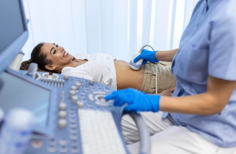
Fetal echocardiography is a non-invasive ultrasound test that uses sound waves to create detailed images of the fetal heart. This includes evaluating the structure, rhythm, valves, and blood flow patterns of the developing heart. Unlike a standard ultrasound, it offers a focused and comprehensive view of cardiac anatomy and function.
When Is It Done?
Fetal echocardiography is usually performed between 18 and 24 weeks of pregnancy. This is when the baby’s heart has developed enough to visualize its anatomy clearly, and there is still enough time left in the pregnancy to plan appropriate care if any abnormalities are detected.
In some high-risk cases, your doctor may recommend an early fetal echo as soon as 16 weeks.
Who Should Consider Fetal Echocardiography?
Not every pregnancy requires a fetal echo, but it is strongly recommended in certain situations, such as:
- Family history of congenital heart disease
- Abnormal findings on a routine ultrasound
- Maternal conditions like diabetes, lupus, or infections (e.g., rubella)
- Use of certain medications during pregnancy (such as anti-epileptics or SSRIs)
- Increased nuchal translucency seen in first-trimester screening
- Multiple gestations (twins or more)
- Assisted reproduction techniques (IVF pregnancies)
Why Is It Important?
- Early Detection of Heart Defects
Fetal echocardiography can identify congenital heart defects such as septal defects, hypoplastic heart syndromes, valve abnormalities, and outflow tract issues. Many of these can be managed or even treated postnatally if diagnosed in time. - Better Planning for Delivery and Care
Knowing about a heart defect in advance allows your medical team to plan for specialized care at birth. This may include choosing a delivery center with pediatric cardiology and neonatal intensive care services. - Reduced Anxiety and Informed Decision-Making
For families at risk, a clear scan can provide peace of mind. If an issue is detected, early counseling helps parents prepare emotionally and practically. - Opportunities for In-Utero Monitoring or Intervention
In rare cases, close monitoring or even in-utero treatment can be considered for certain fetal cardiac conditions.
Expert Care Matters
Fetal echocardiography should always be conducted by a trained fetal medicine or pediatric cardiology specialist for accurate diagnosis and management. Dr. Mamta Phogat, the best Maternal Fetal Medicine Specialist in Faridabad, offers advanced fetal heart screenings and compassionate care tailored to your pregnancy needs. Her expertise ensures early detection, appropriate planning, and the best possible outcome for you and your baby.
Your baby’s heart may be small, but it holds a world of information. Don’t miss the chance to ensure it’s beating just right—from the very beginning.